
Teach lesson
Plant tissues: monocot/dicot stems under the microscope
Students use the Plant Tissues remote lab to compare monocot and herbaceous dicot stems, identify vascular bundles, and defend a classification with real microscope evidence.
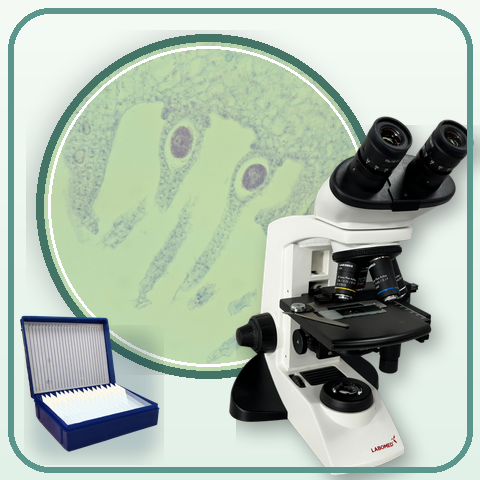
Learning Outcomes
Open the Plant Tissues lab and select the Agricultural Botany collection.
Compare a monocot stem and an herbaceous dicot stem at 4x, 10x, and 40x.
Describe vascular-bundle arrangement and pith/center evidence without inventing measurements.
Recognize xylem, phloem, and parenchyma in a vascular bundle from visible features.
Classify monocot/dicot stems using visual evidence and a real limitation.
Write a CER conclusion suitable for a high-school biology practical.
Student activity preview
Activity Content
Preview only. In a class session, students can fill in responses and submit their work to the teacher.
Predict before observing
7 min
You will compare two real stem cross-sections labelled in the lab as a monocot stem and an herbaceous dicot stem. Your job is not to guess from the label or force the image to match a perfect textbook diagram. Your job is to decide whether the visible vascular-bundle evidence supports each label.
A monocot is a plant with one seed leaf; in many stems, its vascular bundles appear as many small groups. A dicot has two seed leaves; in many herbaceous dicot stems, vascular bundles are arranged near a ring. A stem cross-section is a thin slice cut across the stem, like looking at a stem "round." In that slice, the epidermis is the protective outer layer, the cortex is tissue under the epidermis toward the edge, and the pith is the central region of the stem, often broad in herbaceous dicots. In a real micrograph, those boundaries may look irregular.
A vascular bundle contains:
- xylem, with larger vessels that carry water;
- phloem, with smaller cells that carry sugars;
- surrounding tissue, especially parenchyma.
This activity uses samples 1 and 2 in Agricultural Botany. The other samples are teacher-led extensions.
What to observe before classifying
Use this figure to orient yourself before opening the lab. Your answer must be based on what you observe in your own session, not on copying the support image.
Before observing, which evidence would be most useful for checking whether a stem label is supported by the image?
Explain why vascular-bundle arrangement can help classify a stem. Use at least two of these terms: xylem, phloem, parenchyma, pith, ring, vascular bundles.
Open the lab with a controlled route
10 min
This activity uses a specific lab route: Agricultural Botany, samples 1 and 2, and the 4x, 10x, and 40x objectives. If you cannot select that route, stop and tell your teacher instead of inventing observations.
Follow the route in order: overall pattern first, tissue detail second.
From observation to conclusion
The sequence helps organize the work: describe first, compare next, and finally justify your conclusion with visible evidence.
Open Plant Tissues
Open the lab from TEACH.
If the introduction appears, continue to configuration.
In the collection, select Agricultural Botany.
On the preparation screen, choose slide 1 for Monocot stem cross-section. If a handling warning covers the box, click once on the preparation area to dismiss it, open the sample box, choose slide 1, and then use Skip preparation if your teacher allows it.
Observe the monocot stem at 4x, then use the microscope objective selector to move to 10x. The field/replicate loaded by the lab may vary between sessions; record what you see without trying to force one exact field.
To change sample, use Change sample to return to preparation, open the sample box again, choose slide 2 for Herbaceous dicot stem cross-section, and then use Skip preparation if allowed. Confirm that the observation screen names the dicot sample before recording data.
Observe the dicot stem at 4x and 10x.
For tissue detail, use the objective selector to reach 40x on a vascular-bundle area. If a button such as Look closely does not change the magnification, use the objective control that shows 4x/10x/40x.
If the lab only shows General Botany or blocks the route above, stop and tell your teacher. Do not invent monocot/dicot data.
What is the required route for this activity?
Only answer this if something did not work. Write the exact point where you were blocked: collection, slide 1, slide 2, 10x, 40x, or another control.
Compare the overall stem pattern
15 min
Use 4x to see the whole section and 10x to confirm the pattern near the edge. Observe the monocot stem first, then the herbaceous dicot stem.
Real stem comparison
Use this figure as a search guide. Your graded evidence comes from what you record in your own lab session.
Vascular-bundle pattern
Complete two rows, one per stem. Use 4x for the whole section and 10x to confirm the pattern. In confidence, write high/medium/low plus a reason. In limitation, name what made the view uncertain: focus, crop, brightness, scale, field choice, or access.
| Stem observed | Zoom used for pattern | Bundle arrangement | Center/pith | Confidence | Observation limitation |
|---|---|---|---|---|---|
Which comparison best describes the real images in this practical?
Cite two pieces of evidence from your table that support the difference between the stems. Include at least one point about bundle arrangement and one about the center or pith.
Look inside a vascular bundle
15 min
Now use 40x to inspect a vascular bundle. You do not need exact measurements. Describe tissues from visible features: large vessels, smaller cells, large surrounding cells, and position toward the inside or outside.
Bundle detail
The labels are a search guide. In your own answer, justify from what is visible in your sample.
Tissue detail at 40x
Complete two rows. Use phrases such as 'large vessel-like cells', 'smaller cells next to the large vessels or toward the outside of the bundle', and 'large surrounding filler cells'. If you cannot use 40x, record the blocker and use the best available view, but tell your teacher that the full activity could not be completed.
| Stem | Detail zoom | Xylem evidence | Phloem evidence | Parenchyma evidence | Limitation |
|---|---|---|---|---|---|
Choose one of the two stems and describe the inside of one vascular bundle. Include xylem, phloem, parenchyma, and one limitation of your image.
Which statement best avoids over-interpreting the slide?
Annotated sketch or screenshot
If image upload is slow or confusing, write a structured description first. You may also submit a sketch or annotated screenshot. In any format, include sample, magnification, at least one vascular bundle, and one limitation.
Classify with evidence
8 min
You now have both the overall pattern and the tissue detail. Turn your observations into a defensible classification.
Classify both stems. For each one, write:
- classification: monocot or dicot;
- evidence about vascular-bundle arrangement;
- evidence about center/pith;
- confidence level.
Compare your initial prediction with what you observed. Write one idea you confirmed, one idea you had to revise, and one doubt or limitation you still have.
CER conclusion
5 min
Your conclusion should let a biology teacher see quickly whether you used real evidence rather than memory.
Write a CER conclusion:
- Claim: the main difference you observed between the two stems.
- Evidence: three pieces of evidence from your tables. Include at least one 4x/10x pattern evidence point; if you completed the detail phase, also include one 40x evidence point.
- Reasoning: why that evidence supports monocot/dicot classification.
- Limitation: one real uncertainty in the practical.
Extension: compare other plant samples
15 min
If time and full access allow, each group can choose another Agricultural Botany sample and add one comparison row. The goal is not to memorize 17 preparations, but to ask a reasonable anatomy question.
Class comparison of plant samples
Each group contributes a sample, useful magnification, visible feature, and follow-up question.
| Group | Sample | Useful zoom | Visible feature | Follow-up question |
|---|---|---|---|---|
Choose one additional sample from the confirmed-samples figure. What feature would you like to compare with the stems, and why?